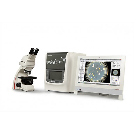
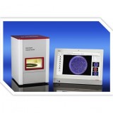
Microscopic cells Analysis / Colony Count / Filter / Inhibition zone measuring spectrometer
CA-MF5
- Remove this product from my favorite's list.
- Add this product to my list of favorites.
Product info
Main Function and Technical Index
1. Colony and inhibition zone digital imaging
1.1. Light source
- Visible light: Highlight the tri-color LED structured light
- 254nm UV: for cavity disinfection, UV mutagenesis
- 366nm UV: excitation Escherichia coli, coliform fluorescent green fluorescent protein
1.2. Optical and lighting control
- closed black box: the elimination of stray ambient light interference
- upper light source: episodic 360 ° flexible shadowless lighting
- lower light source: floating crystal sharp dark-field illumination
- upper light source, lower light source, double light, ultraviolet, free to switch
adjustable color temperature (3500K-8500K), adjustable light intensity
1.3. Photoelectric conversion
- HD Industrial fixed lens: 8mm, 5.0 mega-pixel, 2/3 ", Distortion <-1%, C-Mount
- Room temperature sensitivity CCD camera: 2/3 "color SONY CCD sensor, 1.45 Mega Pixels, C-Mount
2.Colony Counting and Screening
2.1. Basic colony counting function
- Type plate: pouring, coating, membrane filtration, spiral plate, 3M paper, perforated plate
- One key Intelligent Counting (6 mode): Large colonies ,tiny colonies, gray colonies, spreading colonies, certain colonies, multicolor mixed bacteria
- Whole dish colony statistics: total number of colonies statistics, classified display according to 25 gear sizes.
- Selected area statistics: can do statistics with alternative circular, rectangular, arbitrary delineation of regional.
- Diameter classification statistics: set diameter range, statistics specific colony size
- Mouse click statistics: Fast tag, add colonies, suitable for counting colonies at the dish edge
- Colony adhesion segmentation: automatic segmentation colonies adherent to each other, chain colonies can be splitted or not determined by the users.
2.2. Advanced colonies statistics
- Spiral colonies statistics: automatic counting spiral tablet according to FDA standards, support index mode, slow index mode, homogeneous mode ,proportion mode, lawn mode. Compatible with spiral plater of United States SBI, Spain IUL
- Dynamic adjustment statistics: statistics can be dynamically adjusted and corrected, quick access to the best statistical results.
- Estimated deviation statistics: for colonies with many colors and complex cases.
- Level set multiple model algorithm: search operator, get the best image segmentation, adapt to background transformation of the culture medium
- Specific colony statistics: According to the colony color, size, contour feature, identify specific colonies
- Trans statistics: for colony type is extremely complex and medium uniform background
- High adhesion bacteria statistics: for division calculation of multiple adhesion bacteria
2.3. The mesh filter and 3M test piece
- One key statistics for black solid line grid
- 3M total bacteria test piece, 3M S. aureus test piece: one key statistics
- 3M coliform test piece, 3M E. coli / coliform rapid test piece: one key statistics + manual choose
- Bacteria, impurities removed: According to distinguish shape, size, color, automatically remove bacteria, impurities.
- Color classification statistics: according to color accuracy, the proliferation , colony size, contour feature, specific screen colonies
- Multicolor automatic clustering: clustering based on color accuracy, automatically distinguish between 24 kinds of different colors of the colonies
- Specifies multicolor filter: filter 1-8 kinds of colors specified colony at one time
- Transparent Circle Characteristics analysis: Suitable for inhibition zone, hydrolysis ring, color ring, dissolved calcium circle, hemolysis ring, drain ring, dissolved phosphorus loop analysis
- Bi-color ring auto filter
2.5.Description of colonies characterization
- Bacteria, yeast colonies: color, size, shape, surface morphology, edges, gloss, transparency and other features, intelligent descriptions and ordering
- Fungi, actinomycetes colonies: the front color, negative color, size, surface morphology, edge, texture, and other characteristics of intelligent descriptions and ordering
2.6. Microbial Limit Analysis Tools
- Check the applicability of the medium
- Control bacteria examination - colony morphology
- Mold detection: quantitative analysis mildew Ratings
- Series statistics: for complex colonies with uneven medium background
- Parallel statistics: suitable for porous plate, OPKA, SBA analysis
- Clear grid: the elimination of interference of filter grid background
- Manual counting correction: add or delete colonies
- Decontamination areas: the mouse to outline any contaminated areas, automatically remove colonies amount of the contaminated areas
- Eliminate background text: automatically eliminate marker interference
- Background markings removed: automatic elimination of dish stains interference
- Artificial adhesion segmentation: manual division multiple adhesions colonies
- Parameters automatic conversion: diameter of dish, sample dilution input, automatic conversion
- Text, graphics annotation: all kinds of drawing tools and text embedded in English and Chinese
2.9. Calibration and Measurement
- Instrument calibration: The instrument equips with calibration, manual correction calibration
- One-touch rapid measurement: one key measure a large colony ,suitable for single colony analysis of fungi, actinomycetes
- Full dish automatic measurement :whole dish colonies equivalent diameter, area, long and short diameter, perimeter, roundness analysis
- Accurate manual measurement: length, angle, curvature, area, arc, arbitrary curves
3.Inhibition zone measurement and analysis
3.1. Szone inhibition zone multi-mode measuring technology
- Automatic detection: accurate edge detection based on inhibition zone profile, suitable for sharp edges, circular inhibition zone
- Quasidisks approximation: Approximation based on a circular zone of inhibition profile ,fitting for edge cracking, non-circular zone of inhibition
- Manual inspection: click the mouse on the edge of the zone of inhibition by three times into round ,suitable for the fuzzy edge of the zone of inhibition
3.2. Determination of the potency of antibiotics
- A dosage titer assay method: for USP
- Two doses, three doses of law and Consolidate: For Chinese Pharmacopoeia 2010 Edition
- Repeatability self-test: relative error ≤0.01%, repeated accuracy ≤0.002mm
- Homogeneity self-test: relative error ≤0.05%
- Measure difference between platform ≤0.2%
3.3. sulbactam β- lactamase test
- Water verification: According to (A), (B), (D) produced inhibition zone, D-C ≧ 3, B-A ≦ 3, the determined system is established.
- Automatically detects three parallel samples (A), (B), (C), (D) inhibition zone, and data import
- Automatically calculate the average of parallel test, smart discrimination the positive and negative characteristic of the results.
- Automatic alarm for invalid report
4. Database and image processing module
4.1. Image processing and editing
- gradation conversion, image filtering, image segmentation
4.2. Database
- Data storage, intelligent query
- Data Export: Export statistics to Excel table
- Data Security: operator permissions, data modification permission settings
5.Microscopic cell analysis module
5.1. Microscopic imaging
- Microscope: Leica DM500 microscope
- Professional CCD Camera: 1 / 1.8 "color SONY CCD sensor, 5.1 Mega Pixels, C-Mount, Square Housing: 68 × 68 × 43mm
- Resolution: 0.5-1.0 m
5.2. Image display, conversion
- Image display: Real-time dynamic observation, to capture any view image
- Image viewing: rotation, zoom, image conversion, partial observation function
- Image Editing: having images of any area to cut, copy, and paste text input functions
5.3. Microscopic Image Processing
- Adaptive enhancement: The original image and its feature matching resolution enhancement, make the image clearer, more obvious edge in order to observe and recognize the image fine structures.
- Image Adjustment: image brightness, contrast, saturation, RGB three-color any adjustment, grayscale, negative phase diagram of conversion
- Image compensation: linear compensation, the number of ways to compensate for a variety of mathematical, Bell distortion compensation to compensate part of the image to make the image clearer.
- Image sharpening: by enhancing high frequency components of the image, image edge becomes clearer.
- Image formation: by the image formation process, so that a uniform image background.
- Image filtering: Gaussian filter, low pass filtering, median filtering and other six kinds of filtering methods to effectively improve image clarity.
- Edge detection: two detection methods, all three operators with a variety of options to more accurately detect the outline of the extracted image.
- Morphology: erosion, dilation, opening, closing and other nonlinear mathematical morphology.
5.4. Target Measurement
- calibration: online system calibration function for accurate measurement (system built in calibration default value)
- Measurement function: Online measurement of particle diameter, length, curvature, angle, arbitrary curves, the area, etc.
5.5. Particles Statistics
- Automatic statistics: automatic particle counting, and the display area of each particle, circumference, diameter, roundness and other morphological parameters
- Regional statistics: Alternatively rectangular, circular, mushroom and other arbitrary shape regional statistics
- Diameter classification statistics: set diameter range, specific particle size statistics
- Color recognition statistics: according to hue, brightness, saturation, specific particle filter
- Mouse clicks statistics: mouse click to add or remove particles, convenient, fast
- Adhesion segmentation process: according to user needs can be automatically or manually blocking each other split particles
- Variety of statistical algorithms: using a variety of segmentation algorithms for different backgrounds particles Statistics
- Various samples statistics: microscopic image of a plurality of integrated statistics
- Parameters automatic conversion: According to the statistics area of the sample dilution, automatic conversion
5.6. Draw and label
- Drawing: for opened image as needed, draw a straight line, rectangle, circle, and any curves
- Text editor: Open the image of text editing
- Mark: a straight line can be easily and angle dimension
6.Instrument specifications and configuration
- CA-MF5 Multifunctional host
- Leica DM500 microscope, camera switch interface
- Microscope electronic eyepiece (500-megapixel professional CCD)
- Colony analysis software, inhibition zone automatic measurement software, software for measuring the potency of antibiotics, sulbactam β- lactamase test software
High-end integrated compute

